
The ultrasonic diagnostic instrument, commonly known as an ultrasound machine or ultrasound scanner, is a valuable medical device used for diagnostic imaging in various medical specialties. It utilizes high-frequency sound waves to create real-time images of internal body structures, allowing healthcare professionals to visualize and assess organs, tissues, and blood flow. In this article, we will explore the functions and features of the ultrasonic diagnostic instrument, highlighting its importance in medical diagnostics and patient care.
Diagnostic Imaging: The primary function of the ultrasonic diagnostic instrument is to provide diagnostic imaging through the use of ultrasound technology. By emitting high-frequency sound waves into the body, the machine captures echoes as the waves bounce back from different tissues and organs. These echoes are then processed to generate real-time images on a monitor, providing detailed information about the internal structures being examined.
Non-invasive Nature: One of the significant advantages of the ultrasonic diagnostic instrument is its non-invasive nature. Unlike other imaging techniques that may require invasive procedures or ionizing radiation, such as X-rays or CT scans, ultrasound imaging does not involve exposure to harmful radiation or the need for surgical incisions. This makes it a safer option for patients, with minimal discomfort and no known adverse effects associated with its use.
Real-time Imaging: The ultrasonic diagnostic instrument provides real-time imaging capabilities, allowing healthcare professionals to observe and monitor the movement and function of internal structures in real-time. This real-time feature is particularly useful for assessing dynamic processes, such as blood flow through vessels or the movement of a fetus during prenatal examinations. It enables immediate visualization and assessment of structures, aiding in the diagnosis and evaluation of various medical conditions.
Multiple Imaging Modes: The ultrasonic diagnostic instrument offers multiple imaging modes to cater to different diagnostic needs and applications. These modes include:
B-mode (Brightness Mode): The B-mode is the most commonly used imaging mode, producing two-dimensional grayscale images that depict the shape, size, and density of tissues and organs. B-mode imaging provides valuable information for diagnosing conditions in various medical specialties.
Doppler Imaging: Doppler imaging assesses blood flow by detecting changes in the frequency of sound waves reflected by moving red blood cells. It allows healthcare professionals to evaluate blood flow velocity, direction, and abnormalities within blood vessels, aiding in the diagnosis of conditions such as deep vein thrombosis, arterial stenosis, or fetal abnormalities.
Color Doppler Imaging: Color Doppler imaging adds color to the grayscale B-mode images, representing the direction and velocity of blood flow. It provides a visual map of blood flow patterns, enhancing the assessment of vascular conditions and helping to identify areas of reduced or disturbed blood flow.
Spectral Doppler Imaging: Spectral Doppler imaging displays the velocity of blood flow as a graph, allowing healthcare professionals to measure and assess blood flow parameters more accurately. It helps evaluate conditions such as arterial or venous occlusion, valvular dysfunction, or vascular abnormalities.
3D/4D Imaging: Advanced ultrasonic diagnostic instruments offer three-dimensional (3D) and four-dimensional (4D) imaging capabilities. These modes generate three-dimensional images of structures or real-time 3D images, often used in obstetrics for detailed visualization of the fetus. They provide enhanced spatial information and allow for better assessment of complex anatomical structures.
Guided Procedures and Interventions: The ultrasonic diagnostic instrument is utilized for guided procedures and interventions, assisting healthcare professionals in performing precise and targeted procedures. By visualizing internal structures in real-time, the machine helps guide needle insertions, biopsies, fluid aspirations, and other interventional procedures. This improves accuracy, reduces the risk of complications, and enhances patient safety during such procedures.
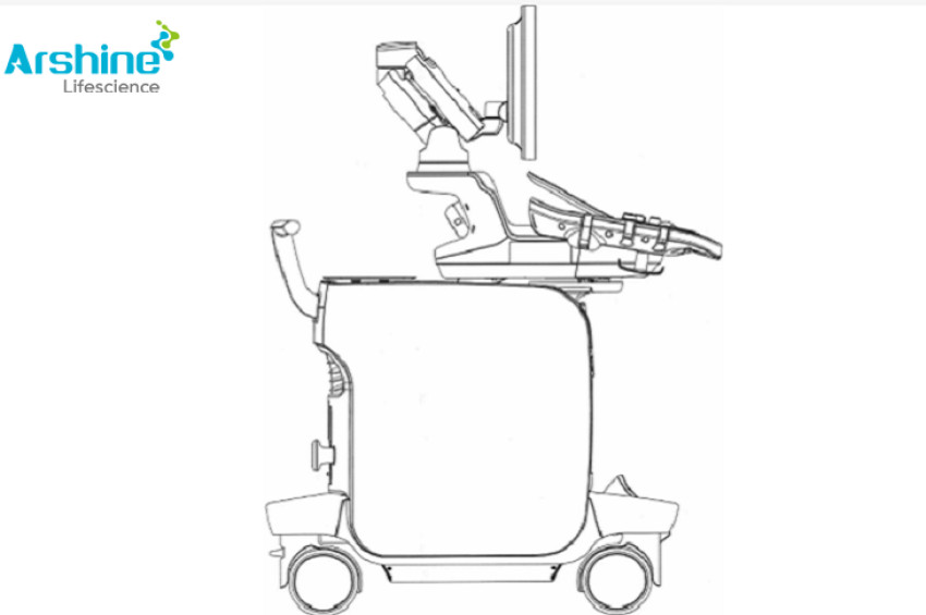
Portability and Point-of-Care Use: With advancements in technology, portable ultrasonic diagnostic instruments have become available, allowing for point-of-care use in various clinical settings. These portable devices are lightweight, compact, and easy to maneuver, making them suitable for bedside examinations, emergency situations, or remote areas where access to traditional imaging facilities may be limited. Portability enables prompt and convenient diagnostic imaging, facilitating timely decision-making and patient care.
Multi-specialty Applications: The ultrasonic diagnostic instrument is widely used across various medical specialties, including but not limited to:
Obstetrics and Gynecology: Ultrasound imaging is commonly used for prenatal examinations, assessing fetal development, detecting abnormalities, and monitoring pregnancies.
Cardiology: Cardiac ultrasound, known as echocardiography, is employed to evaluate the structure and function of the heart, assess cardiac abnormalities, and diagnose heart conditions.
Radiology: Ultrasonography is often used in radiology for abdominal imaging, assessing the liver, gallbladder, kidneys, pancreas, and other abdominal organs. It is also used for musculoskeletal imaging, evaluating tendons, ligaments, and joints.
Vascular Medicine: Ultrasound imaging plays a crucial role in vascular medicine, assessing blood flow, identifying blockages or abnormalities in blood vessels, and guiding vascular interventions.
Emergency Medicine: Portable ultrasound devices are valuable in emergency medicine for rapid assessment of trauma patients, guiding procedures, and aiding in the diagnosis of critical conditions.
Urology: Ultrasound imaging is utilized in urology for evaluating the kidneys, bladder, prostate, and other urological structures, assisting in the diagnosis and management of urological conditions.
Continuous Technological Advancements: The field of ultrasonic diagnostic instruments is continuously evolving, with ongoing technological advancements. These advancements include improved image resolution, enhanced imaging modes, the integration of artificial intelligence (AI) algorithms for automated analysis, and the development of handheld or wearable ultrasound devices. These innovations aim to improve diagnostic accuracy, increase efficiency, and expand the scope of applications in healthcare.
In conclusion, the ultrasonic diagnostic instrument is a versatile and indispensable tool in medical diagnostics. With its non-invasive nature, real-time imaging capabilities, multiple imaging modes, and portability, it enables healthcare professionals to visualize internal structures, assess organ function, guide procedures, and make informed clinical decisions. From obstetrics to cardiology, radiology to emergency medicine, the ultrasonic diagnostic instrument plays a vital role in various medical specialties, contributing to accurate diagnoses, effective treatments, and improved patient care.
https://www.arshinemedical.com/Industry-information/the-function-and-function-of-ultrasonic-diagnostic-instrument